


Osteochondrosis of the lumbar region is a disease that deforms and destroys cartilage tissue of intervertebral discs in the lower back.Without a layer of cartilage, the distance between vertebral is significantly reduced.And with the smallest sharp curves can be switched.The main risk of disease is the possibility of forming an intervertebral hernia.
Can't you lean to pick up the item that fell to the floor?Do you suffer acute pain in lumbar spine and you often go, in the waist in a warm scarf?Don't ignore the condition that bother you.
The osteochondrose of the lumbar region can be returned for a long time.There is no need for the body to be experienced for strength.Love your body.And he'll be back.
The lumbar region makes most of the cargo from all body weight compared to the chest and the storm.Therefore, this subspecies of osteochondrose is the most common.
What are the stages of osteochondrose development?
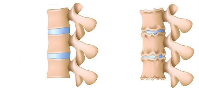
- 1. Phase.Previously.The disk height is reduced.In the fibrous ring (an outer layer of intervertebral disk from the crisp fiber forms a crack. Lumbar muscles are starting to get tired quickly. You feel certain uneasiness in the back.
- 2. Phase.Violations of metabolic processes in the core jacket (central part of the intervertebral disk, which consists of a cartilage jacket): its cells are dead or completely destroyed.Collagen structure (protein structure is based on the connective tissue) of the fibrous rings is also disturbed.Local pain, a person cannot be handled with the physical activity he had previously considered rather feasible.
- 3. Phase.Full destruction of fibrous rings.Neighboring spines stop being stable.Any unpleasant posing causes pain.Due to the experience of the nerve roots that stand away from the spinal cord, the limbs can become less sensitive and mobile.
- Fourth phase.The fabrics of the intervertebral disk becomes cicatric.Vertebra can be shown in a shellfish.Clinical description here depends on individual physiology.
Lumbar pain (lumbago) and pain that provides a knife during a sciatic nerve (Ishias) is one of the most common complaints that patients seek medical attention.Due to the fact that these symptoms are quite frequent in the general population, and their constant growth was also observed, the diagnosis and treatment of such patients will remain one of the main areas of the activities of neurosurgical hospitals.Despite the widespread this pathology, it is necessary to surgically remove the Hernia intervertebral disk (MPD) only in 10% of patients with the clinical picture of Lumbar -algia.In the remaining part of the patients, the best effect has conservative treatment, including drug therapy, physiotherapy exercises, the use of physiotherapy treatment methods, as well as return to previous everyday physical activity.
Stages of the disease
Degenerative-dystrophic processes usually begin by deterioration in the function of absorption of an intervertebral disk strike.
- Deterioration of blood supply to intervertebral disk.In adults, the food of interverters is performed by diffusion: the blood is only on spins, and the discs are powered during dynamic loads (the outflow of the dynamic ingredients, the flow of nutrients). Thus, interventional discsIt is difficult to specifically in the conditions of sitting lifestyle (hypodynamy).
- Changes in the core for pulpic disks.With the decline in blood supply, water supply, sugar and amino acids is disturbed to the nucleop core.Therefore, the production of carbohydrates connecting water.The core is dehydrated, its structure made from the fibrous-like gel, the ability of spring and extinguishing the recording is worsening.This increases the load on the fibrous ring and vertebrae, are more likely to be blocked and injured.
- Changes in the fibrous ring of the intervertebral disk.Due to the flattening of pulposis nucleus, the increased load is located on the disc ring.In the conditions of poor blood supply, the fibrous ring is losing strength.The availability of the spine appears, which can lead to the formation of intervertebral hernia, moving vertebrae and damage to the spinal cord or nerve roots.
- Disk counterpart.Formation of intervertebral hernia.How fibrous ring fibers weakens, for example, the Fulpic core begins, for example, according to the intervertebral channel (disk protrusion).Such astonishing can further lead to rupture of fibrous rings and form a kile.Read more about the process of forming an intervertebral hernia in a separate article - "Efficient treatment of intervertebral hernia at home".
- The spondylosis is destroying intervertebral joints (spondylthrosis), osteophyte growth and environmental ligaments.In parallel with the formation of intervertebral hernia in osteochondrose, they adhere to the damage to intervertebral joints, devastating changes in the vertebrae (cartilage) and ligaments.
As osteochondrosis and development of complications progresses, you must resort to drugs increasingly, increase dose.This leads to high financial costs, as further health deterioration due to the side effects of medications.
The therapy of drugs, as a rule, is supplemented by immobilization of one or friends of the spine using orthopedic corsets of different degrees of rigidity.
Surgical treatment is justified only in cases where the level of spinal arresting is, which confirms the rupture of the MPD in Lumen "in patients with a small protrusion and patient. Method for establishing precise diagnosisis the magnetic resonance (MRI). Approximately 10% of the people of the Joint Population is impossible to run the Routine of Claustrophobia, however, with the appropriate loss of the image obtained. Patients who have previously suffered surgical treatment are needed to show MRI with contrasting armature to delineate.Postoperative scar - changes from the true hernial disk protrusion. In patients with the hernial protuberance of the MPD, when the implementation of the MRI is impossible or the obtained results of non-usual, calculated -Tomographic (CT) milography acquires a special diagnostic value.
Private diagnostic experts who interpret the results of the study, as a rule exaggerate to the degree of disk damage due to the impossibility of comparing clinical data with "finds" during tomography.Conclusions such as "change the patient's age" are almost never found in research protocols.Despite improving neuroimination techniques, the responsibility for a properly deceived diagnosis lies on the shoulders of the clinician, because only he can compare a clinical picture with the data obtained during tomography.Increasing the Tomograma resolution has slightly improved surgical treatment outcomes, but deviations from the norm in asymptomatic patients began to be detected.The process of the process that accompanies the degenerative --distist lesion of the spine has passed serious progress in recent years.Arthropathy of the arched joints widespreadis in the general population and is often discovered in the people of the Central and Old Tenegro during the research of CT.Degenerative changes in the MPD, which are also widely used, are quite discovered, and the MRI is more specific to their diagnosis.At the same time, changes in the MPD are not unusual, not accompanied by a fibrous ring, but are only manifested by a slight "sting" in lumens channels or intervertebral holes.In some cases, degenerative processes that occur in the MPD can lead to the destruction of fibrous rings with subsequent ropes, which causes the migration of the part of the Fulpish-out of the disk with compression of neighboring roots of the spinal cord.The claim that if the pain in the leg is noticed, then it must be harmonized at the roots of the spinal cord is not entirely true.Boilers in the back of the thighs can lead to the back surface of the thighs and the degeneration of MPD and the arched intervertebral joints.For a genuine attack of Ishalgia caused by compression of the Koreshke nerve MPD Hernie, it hurts to radiate on the back surface of the thigh and lower leg.Unspecified pain, limited to the area of gluteal or thighs along the scaling nerve, as well as bilateral pain in glutel areas (either on the left), then are on the left), and then the left-hand side), they are arthropathy of arched joints or diffuse degenerationsMPD.The clinical picture of the Koruska MPD Herninia compression can also be the same pathology (for example, knee joint arthritis).In patients with such pain, surgical treatment will not have an appropriate effect regardless of which pathology will be revealed to the Tomographic exam.In other words, in patients only with a back pain clinic, the removal of MPD hernia will be inefficient, even if tomograms are determined by the MPD protrusion, as usual and occurs.But there are patients in which a typical image of Ishias is accompanied by a pronounced disabled syndrome, while during the studies derived using high perceptiveMographers, the roots of the spinal cord is not determined.This category of patients is inappropriate to perform surgical intervention, because with time, radio culp symptoms, as a rule, lubricated.
It is necessary to clearly imagine mechanisms leading to the development of the Hernian protrusion of the MPD to recommend patients with the volume of permitted movements, not forgetting the work activity.Forces that contribute to the formation of hernial protuberance are the result of degenerative changes in the MPD and reducing vertical (height) and fibrous ring and pulposis nucleus.Sticking fragment of the MPD in 80% shift in the posterior direction, introducing the lumen spinal channel and medial parts of the intervertebral hole.This MPD Hernie shift towards the midline is facilitated by force of the last longitudinal ligament.Up to 10% of hernial relationships are localized laterally and spreads to the intervertal hole (forsin hole) or on the outer edge of the holes in which the cerebrospinal spine exits, which is coming.
In the process of vital activity, dehydration and degenerative changes lead to the loss of the height of the MPD.These pathological processes include a fibrous ring and a Fulpic core.Statable destruction of the core fully against the background of simultaneous degeneration of fibrous rings, as a rule, only leads only the loss of the height of the MPD without significant gatherings.With overcoming changes in fibrous rings, vertical forces affected by the saved Fulpic core and which are their weight, as well as the muscles on the disk, which is excess pressure on the remaining fragment nucleuf for pulposis, which is unable to keep the fibrous ring in place.
Summing these two forces leads to increasing the centrifugal pressure on the MPD, which together with the stretch component operates on fiber fibrous rings can lead to rupture and fragments of the fragments of the remaining window for the window.Once the hernital protrusion is formed, and the "redundant" fragment of the Fulp core was outside the fibrous rings, the structure of the MPD becomes stable again [2].As a result of the forces that influenced the degenerative altered core and fibrous ring of the MPD, they are balanced, and their vector, which contributes to the further protrusion of the core fragments, pale.In some cases, partial degenerative changes in pulpus nucleus contribute to the formation of gas within the MPD, followed by excessive pressure on the remaining fragment.The formation of a herninia is accompanied by the process of forming gas inside the disc.
Excessive and sharp physical activity shown on the patient's backs, on the background of the existing spine, usually is just a trigger that leads to a detailed clinical picture of compression radicular syndrome, which often considered patients, like Primordi Lumbar-Lyun.Clinically, MPD hernia can be manifested with reflex and compression syndromes.Syndrome relate to compression, in which the herself is pulled, clenched and deformed, blood vessels or spinal cords are compressed and deformed.Reflex reflexes include syndrome caused by the effects of the receptors of these structures, mostly the end of return vertebraees, leading to the development of reflex and tonic disorders that are manifested by vasomotor, dystrophic, myofascium disorders.
As mentioned above, surgical treatment of the degenerative-unstrophic lesion of the Essentials are recommended only in 10% of patients, the remaining 90% respond well to conservative measures.The basic principles of using the latter are:
- Relief pain syndrome;
- Reconstruction of the correct position for maintaining the capability of the modified MPD;
- Elimination of muscle and tonic disorders;
- Reconstruction of blood circulation in roots and spinal cord;
- Normalization of conductivity in nerve fibers;
- Elimination of cicatrical and spacing;
- Relocation of psycho-tool disorders.
Treatment
Today, the drugs of the following groups are used in the treatment of osteochondrose and its complications:
- Neto -Ire anti -inflamatory medications (NSAID) - in the form of tablets or drug injections.These funds have the ability to reduce pain, reduce inflammation activity.However, the effect of their use does not last long - from a few hours to two to three days.Therefore, such funds must last for a long time - weeks, and sometimes months.At the same time, these medications negatively affect the mucosa of the gastrointestinal tract.Their long-term reception is full of gastritis development, ulcerative lesions.In addition, they can negatively affect the work of kidney, liver and contribute to the development of hypertension.And, at the same time, these funds do not contribute to cleaning discs from dead cells.Therefore, their use is just a way to relieve symptoms for a while, but not to eliminate the main problem.
- CTEPOID (Gopmonal) anti -infalmator drugs.As a rule, they are used for heavy and impenetrable pain that accompany Kiler, radiculitis, etc.Gopmone have the opportunity to remove inflammation manifestations (due to the oppression of the immune system), alleviate pain.But they also negatively affect the mucous membranes of the stomach and intestines, promote the rinsing of calcium from the bones, inhibit the production of own gopmons.And do not contribute to cleaning the focus of dead cells.
- Papasmolics are drugs that affect muscles or nerves that go into muscle and cause relaxing skeletal muscles.This means helping to release muscle clamps for a while, reduce pain and improve blood flow.But at the same time, they do not help clean the tissues from dead cells.Therefore do not contribute to the healing for osteochondrose.
- Epiduppal blockade - Introduction of hospital and gopmonal funds in the space between the solid brain and peristeum covering the vertebra.As a rule, it is used, for intensive pain - in an acute period of intervertebral hernia, with severe radiculitis, Ishias.Depending on the composition, such injection helps mitigate pain in a period of several hours to a few days.After the expiration date, the disease manifestation returns, as the procedure does not help restore metabolic processes on disks.In addition, when performed, there is a risk of violations of blood vessels and nerves.
Conservative treatments include various orthopedic effects in spine (petty artist, physiotherapy), physiotherapy exercises, acupuncture, drugs. The treatment of the spine should be complex and abolition. As a rule, generalThe principle of conservative measures is the appointment of analgesics, not -steroidal anti -infalm raters (NSAIDs), muscle relaxants and physiotherapy.
The analgesic effect is achieved by the appointment of Diclofenac, Ketoprofen, Lornoxic, tramadol.The pronounced analgesic and antinamental effect has loroxes, which exist in the forms of injection and tablets.
NSAIDs are the most commonly used drugs for degenerative-outstrophistic damage to the spine.They have anti -infalmatory, analgesic and antipyretic effect associated with the combating of the enzymatic cycloking (COC -1 and TSOS -2), which regulates the transformation of arachidic acid in prostaglandins, prostaglands, prostaglands, prostaglands.In older and patients with risk factors for side effects, it is recommended to perform the "lid" of the gastricoter under the "lid".In such patients, after the end of the NSAID injection, the transition to the forms of the COO -2 inhibitor pills, which have the severity of the side effects from the gastrointestinal tract.
To remove pain associated with an increase in muscle tone, it is advisable to include central Musclexants in complex therapy.
The surgical treatment of the degenerative-semi-solor spine lesion is justified by the inefficiency of complex conservative measures (within 2-3 weeks) in patients with a kilical MPD (usually more than 10 mm) and non-intointing radio culinary symptoms.There are urgent markings for surgical intervention with a "discharged" sequence in Luman's spinal channel and expressed compression of the root of the spinal cord.Development of caudal syndrome makes it easier to acute radiculomylomy, which leads to severe hyperalgic syndrome, when even a recipe for drug analgesics, use blockage (with glucocortico and anesthetics) does not reduce pain seriousness.It is important to note that the absolute size of the hernia disk does not have a decisive value for the final decision on surgical intervention and should be considered regarding the clinical picture and find the reveled tomograph exam.In 95% of cases, open access to the vertebra channel is used in hernia.Various chain techniques (coagulation of cold, laser reconstruction, etc.) are currently not currently not present, and their use is justified only for MPD protrusions.Classic open microsurgical removal of the disk of the disk is performed using micro-girger tools, bonnoid bindings or operating microscopes.Analysis of remote treatment results (within 2 years) amounted to the MPD Hernini, which was performed by relapse (27.8% more than 3.5%) in patients who just removed the sequester. The quality of life is more reduced in patients who have pain syndrome, whileRepeat the cylinder formation does not appear clinically.
In conclusion, I would again emphasize the need for a fundamental clinical examination and the analysis of tomograms for the optimal decision on choosing a tactic for the treatment of a particular patient.